Manual treatment, staining, and processing of spheroids and organoids is typically labor-intensive and prone to disruption or loss of samples.
Here we report on use of our microfluidic-based Pu•MA System™ 3D to perform automated assays with single spheroids and populations of organoids followed by a metabolomic profile of spheroids with and without treatment of compounds and immunostainings. The automated pipetting steps allow high reproducibility of results and saves expensive reagents or antibodies at the same time. Different types of plate holders are available for individual assay or analysis requirements.
Spheroid Toxicity & functional assays
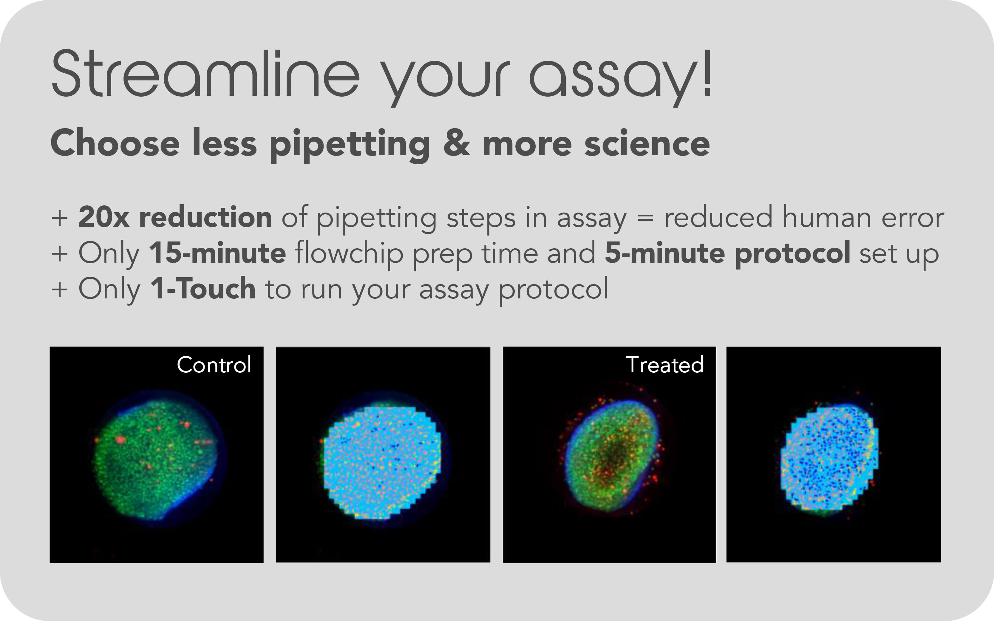
Single spheroids or spheroid populations can be incubated with and without compounds for several days, and then analyzed with different parameters. Collecting supernatant in combination with cell viability assays or stainings are possible for single spheroids or population of structures. Low attachment flow chips allow long-term cultivation with automated media changes and/or treatment of the cells or co-cultivation with immune cells. Assays can be measured and analyzed direct in the flow chip, due to compatibility with plate readers in 384-well format.
Interested? Learn more about this in our free App Note "Pu•MA System™ 3D for Spheroid Toxicity Testing and In Situ Imaging"
Download
Single Spheroid Metabolomics
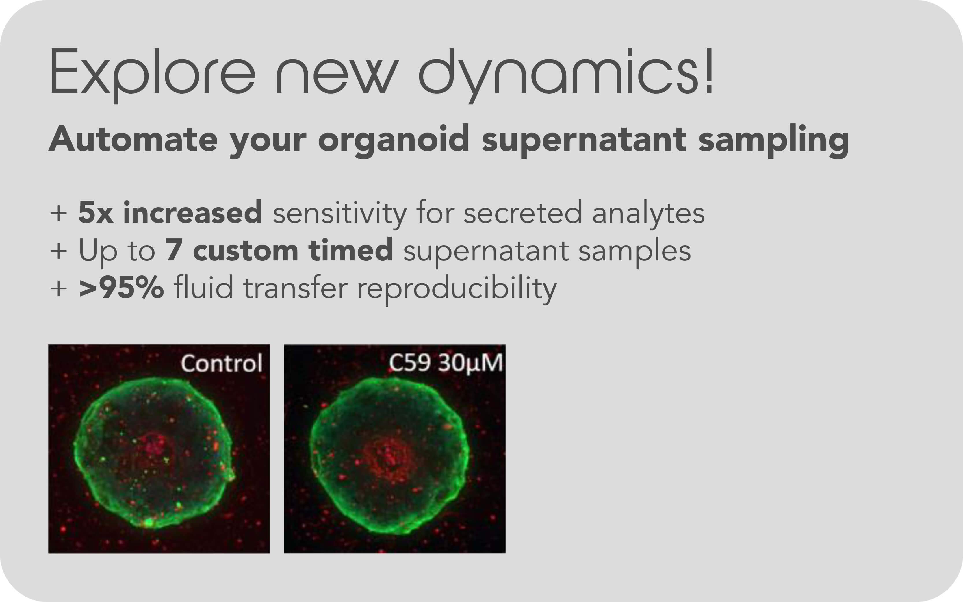
Use of Pu•MA System™3D for automated media and compound treatment along with in situ lysing of spheroids for metabolomics. The ability to lyse organoids in situ in order to capture metabolomic profiles with minimal disruption of the spheroids shows great promise for research. All reagents are loaded into flow chips and then incubation, media exchanges, cell secretion sampling and other steps are executed by the system program with a recovery rate of up to 95% without drying cells.
Interested? Learn more about this in our free App Note "Use of Pu•MA System™ 3D for Single Spheroid Assays with Downstream Metabolomics"
Download
Immunofluorescence staining in ECM
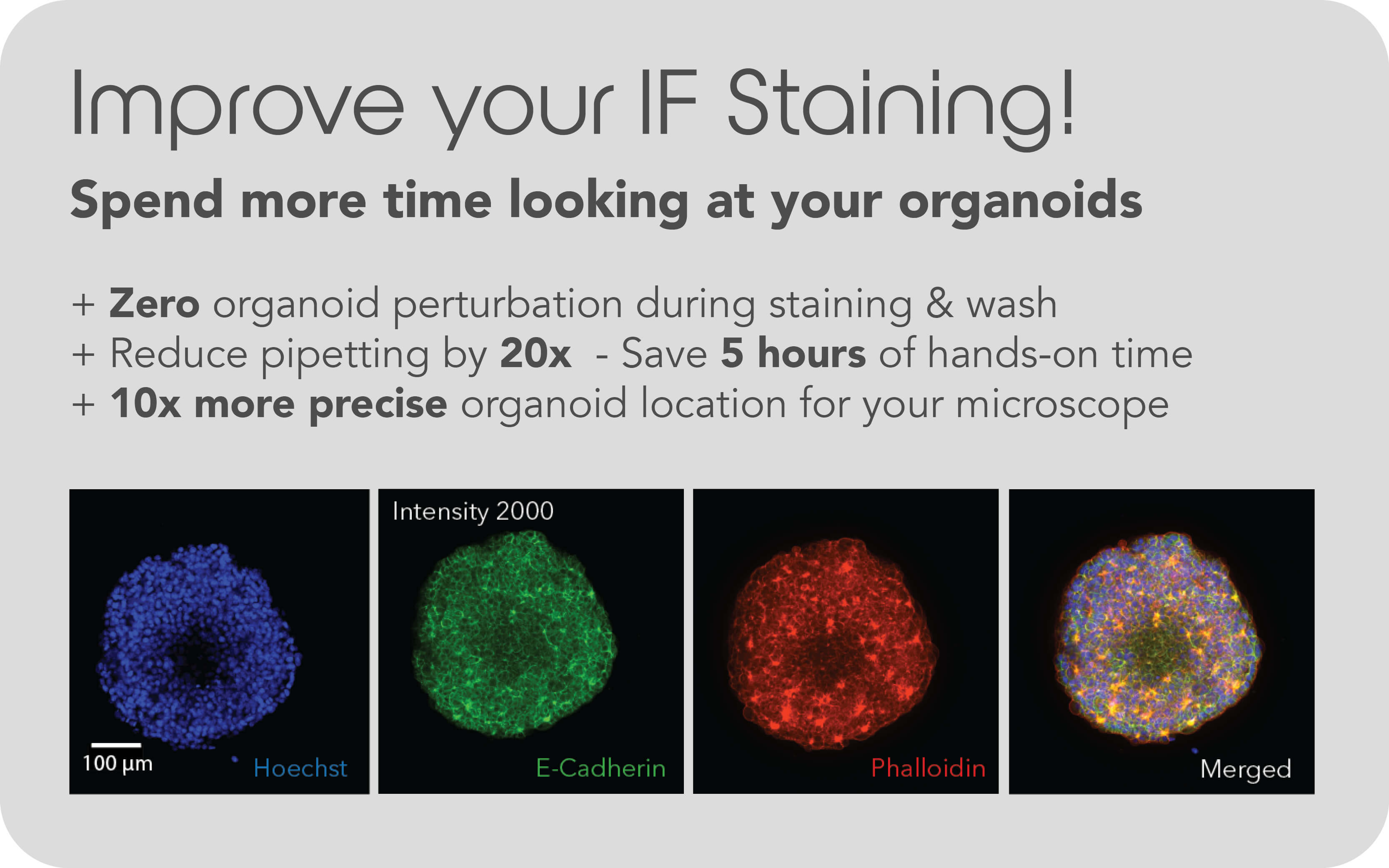
Immunofluorescence staining (IF) is a widely used method to identify and quantify biomarker expressions and their cellular localizations. One of the challenges faced by researchers is that IF staining of delicate spheroids/organoids tend to tedious if done manual, due to loss of samples or disruption. Automated IF staining for biomarker detection is working with different types of ECM and cell types like cell lines spheroids and patient derived tumoroids growing in natural or engineered ECM.
Interested? Learn more about this in our free App Note "Automated Biomarker Identification using IF Staining using the Pu•MA System™ followed by High-Content Confocal Imaging"
Download
Immuno-Oncology & Immune Cell-Co-Culture
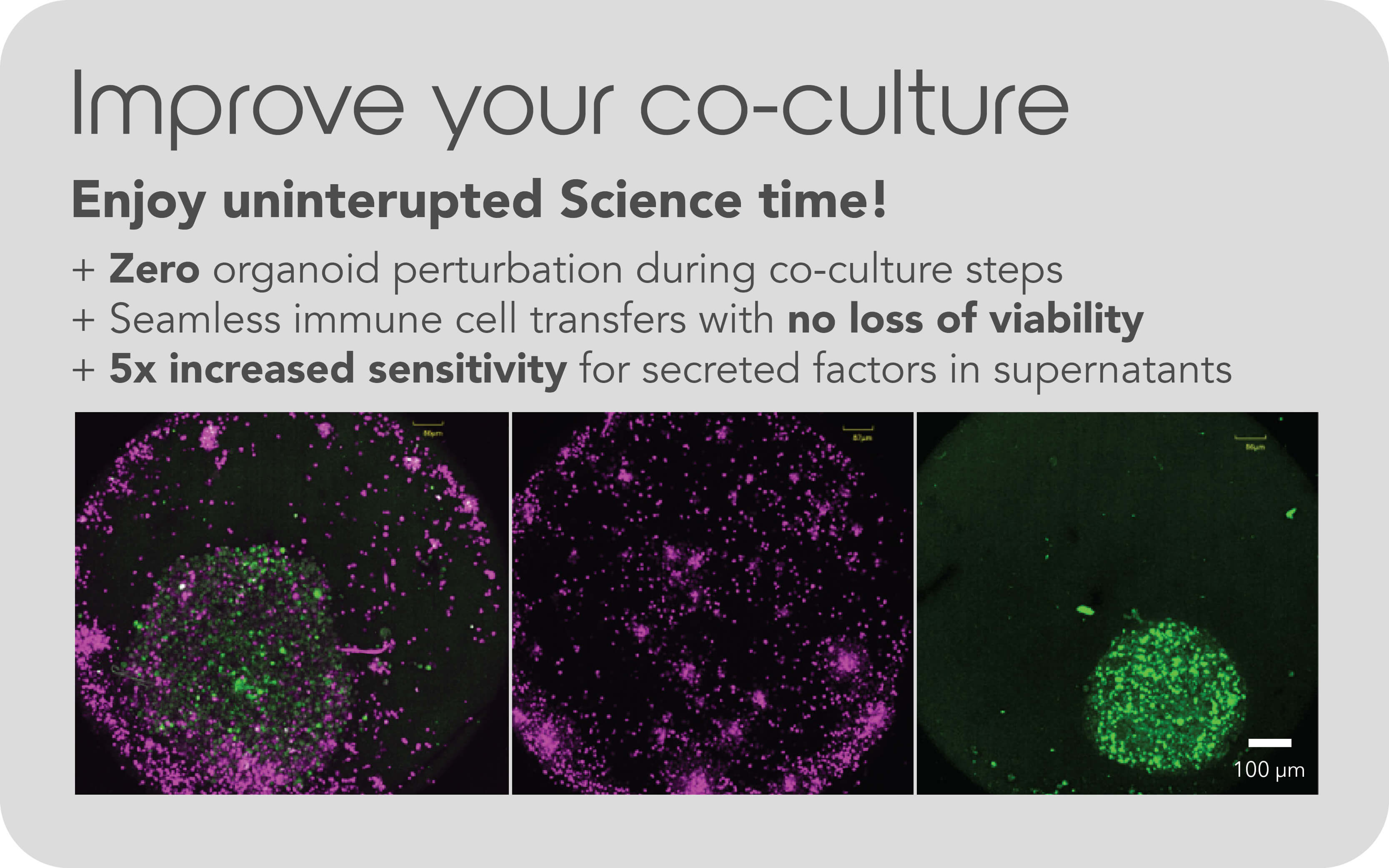
T-cell therapy or chimeric antigen receptor T-cell therapy (CAR-T) is becoming an important approach for anti-cancer treatments. Therefore, researchers are continually discovering new targets and mechanisms to improve the understanding of the complex tumor microenvironment. The use of the Pu•MA System allows co-culturing without disturbing the organoids and collect supernatant to analyze secreted factors. It is possible, to include staining steps for imaging the co-cultures in the automated workflow.
Sounds exciting? Learn more in our free App Note "Automated 3D Co-culture Assays Using Activated T-cells Within the Pu·MA System"
Download
Webinar
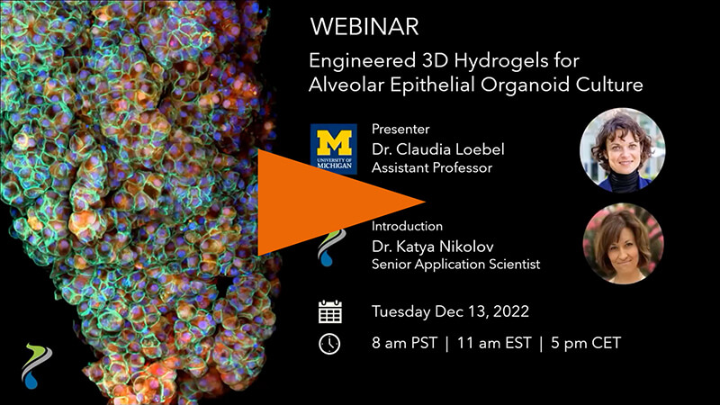
Webinar on demand: Learn how you can perform automated organoid assays with ECM
In this webinar, Professor Dr. Claudia Loebel (University of Michigan) presented her latest research on engineered hydrogels with focus on the unique technology she is developing to study alveolar epithelial organoids and the application of both in the study of pulmonary diseases.
Watch now
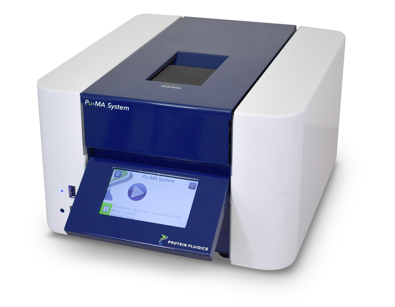
Automated 3D cell-based assays with the Pu•MA System™ 3D
The Pu•MA System™ has been designed to automate assays for spheroids or organoids by maintaining the 3D cell model in a special desigend flowchip to allow automated media or reagent exchanges.
- Automated workflow for complex assay protocols
- Protected sample chamber to prevent cell damage
- Automatic execution of media change, compound additions and supernatant sampling
- Small well with low reagent volume
- Compatible with high content imaging and plate reader systems
- Small footprint fits on bench and incubator
Coming soon: Pu•MA System EC
with temperature control (Ambient + 5 °C up to 40 °C +/- 0.5 °C), CO2 (0-10%, +/- 0.1%) and humidity (~95% RH with use of water reservoir holders)